BREAST Acellular Dermal Matrices in Primary Breast Reconstruction: Principles, Concepts, and Indications Maurice Y. Nahabedian, M.D. Washington, D.C.
Summary: Prosthetic breast reconstruction using acellular dermal matrix is currently used by many plastic surgeons. As our understanding of these matrices expands, our results and outcomes are becoming more reproducible and predictable. As with most new technologies, there is a learning curve associated with using acellular dermal matrix. There are principles and concepts that should be heeded when considering their use. The purpose of this article is to review some of the important principles and concepts to improve our understanding of how these matrices perform and what can be expected of them. (Plast. Reconstr. Surg. 130 (Suppl. 2): 44S, 2012.)
B
reast reconstruction following mastectomy continues to remain a topic of interest in plastic surgery. Over the past several years, there has been a paradigm shift in the traditional options and techniques available for breast reconstruction because of various advancements and innovations. As a result, the number of women choosing breast reconstruction has increased not only because of the expanded options but also because surgical outcomes have improved. Breast surgeons are now able to safely and effectively perform skin-sparing and occasionally nipple-areola–sparing mastectomy to preserve the natural skin envelope of the breast. Reconstructive surgeons are now able offer autologous tissue options using perforator flaps such as the deep inferior epigastric perforator, superficial inferior epigastric artery, and superior gluteal artery perforator flaps that preserve the donor-site muscles. Prosthetic devices are now available using shaped devices and cohesive silicone gels. All of these innovations and advancements have served to provide women with safer and more effective options when considering reconstruction following mastectomy. The innovation that arguably has had the most significant impact in recent years is the use of acellular dermal matrix in the setting of prosthetic reconstruction.1–7 The primary reason for this is From the Department of Plastic Surgery, Georgetown University. Received for publication February 2, 2012; accepted March 19, 2012. Copyright ©2012 by the American Society of Plastic Surgeons DOI: 10.1097/PRS.0b013e31825f2215
44S
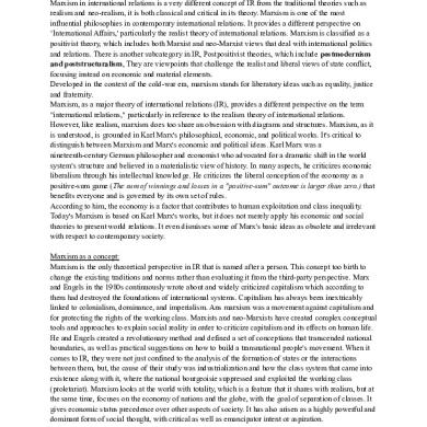
that acellular dermal matrix provides additional control over the mastectomy space and the prosthetic device and creates the potential for improved surgical outcomes. It enables surgeons to optimally position a tissue expander or implant on the chest wall, facilitating one- and two-stage reconstruction. Many of these matrices have regenerative potential and are capable of revascularization, recellularization, and providing tissue support (Fig. 1). Although acellular dermal matrix provides another tool for the plastic surgeon, there is a learning curve that must be appreciated to obtain desired results. The purpose of this article is to review relevant concepts, principles, and indications associated with using acellular dermal matrix. It is important to remember that there are many different acellular dermal matrix materials currently available. A PubMed literature search will identify dozens of articles regarding acellular dermal matrix and breast reconstruction. Most of these articles are level 3, 4, and 5 in nature, with a paucity of level 1 and 2 articles. What becomes
Disclosure: Dr. Nahabedian serves on the speakers bureau and is a consultant for LifeCell Corporation (Branchburg, N.J.). He receives honoraria for speaking about the various LifeCell products, which include AlloDerm, Strattice, and Spy fluorescent angiography in the setting of autologous and prosthetic breast reconstruction. No financial or administrative support was received for the preparation of this article.
www.PRSJournal.com
Volume 130, Number 5S-2 • Primary Breast Reconstruction
Fig. 1. The typical appearance of revascularized AlloDerm at the time of device exchange is illustrated.
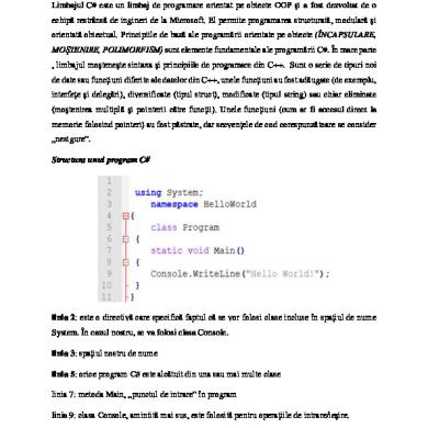
readily obvious is that most of the articles are based on the techniques and outcomes of a single product (Fig. 2). The content of this review will be based on the published literature and on personal experience using acellular dermal matrix for prosthetic breast reconstruction.
INDICATIONS FOR ACELLULAR DERMAL MATRIX IN PRIMARY BREAST RECONSTRUCTION Acellular dermal matrix can be used in most patients who are candidates for prosthetic reconstruction.1–7 Personal experience has demonstrated success in women with a variety of breast volumes that range from A to D cup size. It can be used in young and elderly patients. Maximal ben-
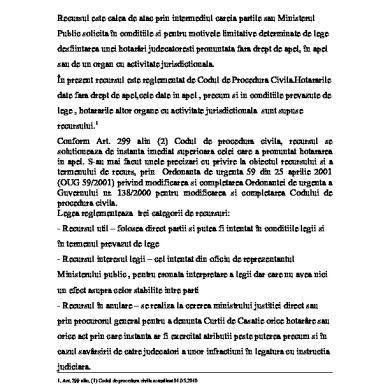
efit of acellular dermal matrix is obtained when it is used in the setting of immediate breast reconstruction (Fig. 3); however, it can also be used in the setting of delayed breast reconstruction in women who have some degree of skin laxity (Fig. 4). With delayed breast reconstruction, the technique involves recreating the mastectomy defect, elevating the pectoralis major muscle, and securing the acellular dermal matrix in the standard fashion (Fig. 5). Given that the incidence of bilateral breast reconstruction is increasing, there are fewer issues related to symmetry and patient satisfaction is high. There are, however, certain patients in whom acellular dermal matrix is less effective. These include those who are morbidly obese, have had a prior mastectomy and radiation, have severely compromised vascularity to the skin flaps immediately following mastectomy, and are actively using tobacco products. In these complex patients, the risks associated with prosthetic reconstruction may outweigh the benefits. The reasons to avoid the use of acellular dermal matrix in these patients warrant explanation. Morbidly obese patients, defined as those patients with a body mass index greater than 40, do not generally fare as well as patients with a lower body mass index from aesthetic and functional perspectives.8 With morbidly obese patients, the thickness of the surrounding subcutaneous layer can be in excess of 3 cm. Thus, the device sits deep in the mastectomy space and is unable to generate any significant projection even when fully expanded. In addition, the thickness of the mastec-
Fig. 2. The number of published peer reviewed manuscripts as listed on PubMed is tabulated based on the specific acellular dermal matrix. (PubMed was accessed January 20, 2012.)
45S
Plastic and Reconstructive Surgery • November Supplement 2012
Fig. 3. The typical appearance of two-stage prosthetic reconstruction using AlloDerm as a pectoralis major extender.
Fig. 4. Preoperative view of a patient scheduled for delayed prosthetic breast reconstruction using AlloDerm. The patient had received no previous radiation. Note the redundant skin envelope following skin-sparing mastectomy.
tomy skin flaps is sometimes in excess of 1 to 2 cm. It has been a personal observation that acellular dermral matrix adherence becomes less predictable when the thickness of the mastectomy skin flaps exceeds 1 cm. This may be because as the distance from fat surface to the subdermal plexus increases, the surface fat (distal) is less vascularized than deeper fat (proximal) to the subdermal plexus and may be prone to nonadherence. The effects of radiation therapy on mastectomy skin site are well known and include increased fibrosis, decreased elasticity, and compromised vascularity.9 Breast skin and gland that have been previously radiated can pose reconstructive challenges. Delayed prosthetic reconstruction following mastectomy and radiation is limited be-
46S
Fig. 5. An intraoperative view after delayed prosthetic breast reconstruction using AlloDerm.
cause of the relative inability of the skin to adequately expand. Although, some acellular dermal matrices have been demonstrated to revascularize in the setting of prior radiation, the decreased soft-tissue elasticity will limit its efficacy. Patients who have had aggressive mastectomy procedures with very thin skin flaps that are borderline viable are also not good candidates for acellular dermal matrix. In these patients, it may be prudent to avoid immediate prosthetic reconstruction with matrix material and merely close the skin, especially when tissue perfusion cannot be assessed. Fluorescent angiography is a useful tool in these situations to assess tissue perfusion.10 If the perfusion is poor, debridement and skin closure are recommended, and delayed reconstruction with or without acellular dermal matrix can be considered following adequate healing. Finally, it is well known that tobacco products will compromise vascularity and increase the incidence of delayed healing, tissue necrosis, and incisional dehiscence.11 It is my policy to encourage all patients to avoid tobacco products 4 weeks before surgery. Failure to comply will result in completion of the mastectomy with delayed reconstruction. Failure to stop using tobacco products can increase the risk of reconstructive failure.
ACELLULAR DERMAL MATRIX FACILITATES PARTIAL MUSCLE COVERAGE TECHNIQUES The ideal position of a prosthetic device following mastectomy is generally agreed to be under the pectoralis major muscle. An ongoing debate, however, is whether the device should be totally or partially under the muscle. In most cases, the mus-
Volume 130, Number 5S-2 • Primary Breast Reconstruction cle includes the pectoralis major only, but in some cases the proximal rectus abdominis and the serratus anterior may be included. It is generally agreed that prepectoral placement of a device following mastectomy may predispose patients to poor cosmetic outcomes or reconstructive failure in the event of thin skin flaps, compromised tissues, or delayed healing, and it is therefore not usually considered. The arguments regarding device placement are diverse. Advocates for total muscle coverage will cite enhanced and more stable coverage as well as fewer complications, such as infection and seroma. Critics of total muscle coverage will cite that there is a greater potential for malposition, less control of the lower pole, increased need to partially elevate surrounding muscles such as the rectus abdominis and serratus anterior muscles, and, finally, less intraoperative expansion. Partial muscle coverage can be performed with or without acellular dermal matrix. The benefit of partial muscle coverage is based on the concept that release of the inferior insertion of the pectoralis major muscle will improve device projection, allow for device positioning along the inframammary fold, and allow for increased fill volume. When partial muscle coverage without acellular dermal matrix is performed, there is an increased risk of rippling and wrinkling along the lower pole, especially when the lower mastectomy skin flap is thin. In addition, there is an increased risk of device exposure in the event of delayed healing. When partial muscle coverage with acellular dermal matrix is performed, the additional layer can theoretically minimize rippling and wrinkling because of the added thickness, serve to better define the natural breast landmarks, provide additional tissue support, and minimize device exposure in the event of delayed healing. These observations are based on personal experience. In essence, the acellular dermal matrix is serving as a tendon, anchoring the pectoralis major muscle to the chest wall at the inframammary fold, thus minimizing “window-shading” or superior displacement of the muscle (Fig. 3). Clinical studies comparing total and partial muscle coverage suggest that both can be effective, albeit with limitations. Sbitany et al. compared 50 women following total muscle coverage and 50 women following partial muscle coverage and placement of acellular dermal matrix.12 Mean intraoperative fill volume was 412 cc with partial muscle coverage and 130 cc with total muscle coverage (p ⫽ 0.0001). The mean number of postoperative expansions was 1.72 with partial and
4.31 with total muscle coverage (p ⫽ 0.0001). Thus it was demonstrated that partial muscle coverage with acellular dermal matrix increased intraoperative expansion and decreased the number of postoperative expansions. The rate of postoperative complications was 18 percent with partial and 14 percent with total muscle coverage (p ⫽ 0.79). Antony et al. compared complications following total muscle coverage in 2910 women with partial muscle coverage and acellular dermal matrix in 153 women.13 The incidences of seroma, infection, reconstructive failure, and exposure were higher in the women following partial compared with total muscle coverage. It was concluded that total muscle coverage resulted in fewer complications. In contrast to the results of the study by Antony et al., Vardanian et al. compared 203 women with partial muscle coverage and acellular dermal matrix to 129 women without acellular dermal matrix (total and partial muscle coverage).14 The acellular dermal matrix cohort had fewer complications compared with the non–acellular dermal matrix cohort (29.3 percent versus 40.3 percent, p ⫽ 0.038). Table 1 summarizes infection following prosthetic reconstruction using acellular dermal matrix in several studies from 2005 to 2010.1,4,5,14,15–19 Aesthetic outcomes following acellular dermal matrix are categorized based on various parameters, including patient satisfaction and the occurrence of untoward events such as capsular contracture, malposition, and rippling. Vardanian et al. evaluated various parameters in their comparative study.14 The capsular contracture rate was 3.8 percent in the acellular dermal matrix cohort compared with 19.4 percent in the non–acellular dermal matrix cohort (p ⫽ 0.002). There was less bottoming out (4.8 percent versus 12.4 percent), rippling (3.8 percent versus 10.9 percent), and Table 1. Recent Studies Comparing Infection Rates Study 15
Marguiles et al. Salzberg4 Zienowicz and Karacaoglu5 Breuing and Colwell1 Preminger et al.16 Nahabedian17 Chun et al.18* Salzberg et al.19 Vardanian et al.14
Year
No. of Breasts
No. of Infections
2005 2006
50 76
2 0
4 0
2007
30
0
0
2007 2008 2009 2010 2011 2011
67 45 100 269/206 466 208
2 3 5 24/10 1 2
Percent
3 6.7 5 8.9/4.9 0.2 1
*In the study by Chun et al., the numerator represents cases associated with primary and secondary infection. The denominator represents cases with primary infection.
47S
Plastic and Reconstructive Surgery • November Supplement 2012 mechanical shift (1.9 percent versus 9.3 percent) in the acellular dermal matrix cohort. Improved patient satisfaction was demonstrated in a case report describing a single-stage bilateral implant reconstruction.20 One breast had total muscle coverage and the other had partial muscle coverage with acellular dermal matrix. There was improved projection, contour, and inframammary fold definition in the breast with partial muscle coverage based on patient reporting (Fig. 6). Although a definitive conclusion cannot be drawn based on a single case, this has been observed in the majority of cases.
ACELLULAR DERMAL MATRIX FACILITATES CONTROL OF THE MASTECTOMY SPACE AND PROSTHETIC DEVICE The important concept to be emphasized in this section is that improved control of the mastectomy space and prosthetic device can lead to improved aesthetic outcomes. This is true for both one-stage and two-stage prosthetic reconstruction. The postmastectomy pocket is usually bordered by the inframammary fold, lateral sternal edge, anterior axillary fold, and the second rib. The footprint or anatomic dimensions of this space are usually different from that of a tissue expander or implant. Traditionally, plastic surgeons have relied on suturing techniques to maintain a postmastectomy pocket of appropriate dimensions.
Fig. 6. A postoperative 2-year follow-up photograph following one-stage total muscle coverage reconstruction on the left and two-stage partial muscle coverage with AlloDerm reconstruction on the right. (Reprinted from Nahabedian MY. Mastectomy, nipple-areola preservation, and immediate implant reconstruction: Are total and partial muscle coverage techniques aesthetically equivalent? Plast Reconstr Surg. 2010;126:319e-320e).
48S
Before using acellular dermal matrix, partial muscle coverage was performed by securing the inferior edge of the pectoralis major muscle by one of two methods.3 The first method was to use marionette sutures to stabilize the position of the pectoral muscle to prevent window shading. The second method was to suture the inferior edge of the pectoralis major muscle to the lower mastectomy skin flap. Although successful in the majority of cases, total control of the lower pole was not always achieved. Sometimes the medial, lateral, and inferior boundaries of the mastectomy space would be excessively undermined, resulting in implant displacement or movement. Thus, the purpose of acellular dermal matrix is to maintain appropriate pocket dimensions, facilitate compartmentalization, and maintain natural breast landmarks. This leads to another important point that relates to selecting the appropriate size of matrix material to use. Acellular dermal matrix for breast reconstruction is available in different sizes: 16 ⫻ 4 cm, 16 ⫻ 6 cm, 16 ⫻ 8, cm, and 20 ⫻ 8 cm. It is also available in variable thicknesses, from less than 1 mm, 1 to 2 mm, and greater than 2 mm. As a novice using acellular dermal matrix for prosthetic reconstruction, the 16 ⫻ 4-cm piece was typically used, as there was no algorithm for acellular dermal matrix selection based on device volume. Unfortunately, the 16 ⫻ 4-cm sheet was too short to permit optimal projection of the tissue expander, because the height of the sheet was too short to extend to the apex of the device. Sheets that were 6 to 8 cm in height would extend to the apex of the device with greater ease and allow for better breast projection. With two-stage reconstruction, there is greater flexibility in the selection of the acellular dermal matrix. Medium to tall sheets (6 to 8 cm) can be used because the device is inserted with minimal volume and filled intraoperatively (Fig. 7). Once sutured in place, the acellular dermal matrix has the potential to stretch.21 With one-stage reconstruction, sheets that are 8 cm in height are typically necessary because the devices are usually prefilled (Fig. 8). The additional height is necessary in order for the matrix material to reach the apex of the device. A sheet that is too short can result in lower pole compression, limited projection, and superior displacement. Thus, an algorithm for acellular dermal matrix selection based on sheet size, thickness, and device volume was created (Table 2). Larger devices typically require larger sheets. Another observation was that thinner sheets tended to adhere to the surrounding tissues with greater predictability. The process of adherence
Volume 130, Number 5S-2 • Primary Breast Reconstruction
Fig. 7. An intraoperative photograph depicting a two-stage prosthetic reconstruction using a tissue expander.
Fig. 8. An intraoperative photograph depicting a one-stage prosthetic reconstruction using a prefilled silicone gel implant.
There are several important principles and concepts regarding the technique of acellular dermal matrix placement as it relates to control of the device. These are based primarily on my technique. A complete review of acellular dermal matrix technique is provided in other sections of this supplement. After elevation of the pectoralis major muscle, the matrix is sutured medially from the inferomedial corner of the pectoralis major to the medial border of the inframammary fold. This vertical suture line is relatively loose to permit medial projection as the device expands. Inferiorly, the acellular dermal matrix is sutured to the fascia at the level of the inframammary fold. An interrupted suturing technique is preferred to permit some degree of communication between the dorsal and ventral acellular dermal matrix compartment. Some surgeons will perforate or “pie-crust” the matrix material to facilitate fluid evacuation and minimize seroma formation. Laterally, the acellular dermal matrix is sutured just under the edge of the device to adequately compartmentalize and prevent lateral migration (Fig. 9). It is important to identify the lateral intercostal nerves and avoid suture ligation that may cause untoward pain. Most studies evaluating the incidence of seroma have demonstrated a slight increase in the setting of acellular dermal matrix. Table 3 includes several studies reporting on the incidence of seroma following prosthetic reconstruction with and without acellular dermal matrix.12,14,16,22 Minimizing the incidence of seroma is dependent
Table 2. Author’s Algorithm for Selection of Acellular Dermal Matrix Size Based on the Volume of the Prosthetic Device Used Volume Range (Prosthetic Device) Sheet Size 16 ⫻ 4 16 ⫻ 6 16 ⫻ 8 20 ⫻ 8
cm cm cm cm
300 to 400 cc
400 to 500 cc
500 to 600 cc
600 to 700 cc
700 to 800 cc
Yes Yes No No
No Yes No No
No No Yes No
No No Yes No
No No No Yes
usually takes approximately 2 to 8 weeks to occur. In the event of poor adherence, the nonadhered portions of acellular dermal matrix are usually removed and the adherent portions are retained. In the event of poor adherence after several weeks, it is unlikely that adherence with that sheet will occur.
Fig. 9. An intraoperative photograph illustrating the lateral edge of the prosthetic device and AlloDerm.
49S
Plastic and Reconstructive Surgery • November Supplement 2012 Table 3. Recent Studies Comparing Seroma Rates Vardanian et al.14 Sbitany and Serletti22 Preminger et al.16 Sbitany et al.12
Study Type
Year
Seroma (ADM Cohort)
Seroma (No ADM Cohort)
Retrospective Metaanalysis Retrospective Retrospective
2011 2011 2008 2009
5/208 (2.4%) 8.40% 3/45 (6.7%) 3/50 (6%)
2/129 (1.56%) 4.30% 2/45 (4.4%) 3/50 (6%)
upon several factors, one of which includes proper drain placement (Fig. 9). This serves to minimize fluid accumulation, provide an environment of negative pressure, and ensure contact between the acellular dermal matrix and mastectomy skin flap. The exact time required for revascularization and recellularization to occur is unknown but appears to be initiated within 1 to 2 weeks based on tissue histology. It has been a personal observation that adherence is facilitated by placing the acellular dermal matrix with the dermal side (not the basement side) toward the mastectomy skin flap. Once the acellular dermal matrix begins to adhere, the likelihood of developing a seroma will diminish. For this reason, the drain between the mastectomy skin and acellular dermal matrix is usually retained for 1 to 2 weeks and is not removed until the 24-hour output is less than 30 cc/day. Clinical studies have validated the concept that acellular dermal matrix will improve surgical outcomes following one- and two-stage prosthetic reconstruction.19,23 Salzberg et al. reviewed their 8-year experience with one-stage prosthetic reconstruction.19 The overall complication in 466 breasts was 3.9 percent and included loss, exposure, or malposition of the implant, as well as hematoma, infection, and capsular contracture. Colwell et al. compared outcomes following one-stage reconstruction with acellular dermal matrix to two-stage reconstruction without acellular dermal matrix.23 The complication rate was similar between the two cohorts (14.8 percent versus 19.6 percent, p ⫽ 0.18).
ACELLULAR DERMAL MATRIX CAN MINIMIZE PERIPROSTHETIC FIBROSIS Several clinical studies have reported that capsular contracture may be minimized in the setting of prosthetic devices and acellular dermal matrix.1,2,4 –7 In these studies, the incidence of capsular contracture ranged from 0 to 2 percent. A limiting factor was the relatively short follow-up, which ranged from 10 to 18 months. Studies with long-term follow-up of 5 years or longer are necessary to make definitive conclusions. In a review of 260 patients and 466 breasts over an 8-year period, Salzberg et al. demonstrated that the in-
50S
cidence of capsular contracture was 0.4 percent at a mean follow-up of 28.9 months (range, 3 to 98 months).19 There have been several experimental and clinical studies that have attempted to explain this phenomenon. In an experimental study in rabbits, Uzunismail et al. implanted silicone sheets into the dorsum of 20 rabbits.24 In 10 rabbits, the sheets were wrapped in acellular dermal matrix, and in the other 10 they were not. All periprosthetic tissues were analyzed at 13 weeks. In the acellular dermal matrix group, the capsule was thin with minimal cellularity or inflammatory infiltrates, whereas in the non–acellular dermal matrix group, the capsule was thick with increased cellularity. In another experimental study in monkeys, Stump et al. observed that implanted devices that were partially wrapped in acellular dermal matrix demonstrated less capsule formation than those devices that were not wrapped in acellular dermal matrix.25 These two experimental studies suggest that capsule formation is minimized in the setting of acellular dermal matrix. In a clinical study, Basu et al. studied the histology of implanted acellular dermal matrix and native capsule in 23 women following prosthetic reconstruction.26 Histologic analysis of the matrix material demonstrated a lack of granulation tissue and vascular proliferation as well as a mild increase in collagen and inflammatory infiltrates. Capsule histologic analysis demonstrated abundant granulation with mild vascular proliferation as well as a moderate increase in collagen and inflammatory infiltrates. This clinical study demonstrates that the presence of acellular dermal matrix seems to attenuate the process of fibrosis in humans. Explanations for this phenomenon are debatable but are most likely related to the inflammatory response. The association of inflammation and fibrosis is well known. Prantl et al. demonstrated that in women with silicone gel implants and capsular contracture, the capsules are characterized by vascular proliferation and the presence of a lymphocytic and mononuclear infiltrate as well as silicone particles.27 In an in vitro study, Orenstein et al. demonstrated that the inflammatory response to AlloDerm is significantly less than
Volume 130, Number 5S-2 • Primary Breast Reconstruction that of all other acellular dermal matrix materials tested.28 On the basis of the observation that periprosthetic fibrosis with acellular dermal matrices may be minimized, some surgeons are advocating prepectoral placement of the device with total anterior coverage using acellular dermal matrix. The rationale is that the device will remain covered in the event of delayed healing and that the operation would be simplified by not having to elevate the pectoralis major muscle. Unfortunately, there are no published reports describing this technique, thus it will not be reviewed in detail. Suffice it to say that this technique is being evaluated. In summary, Basu et al. demonstrated increased inflammation and vascular proliferation in capsule compared with acellular dermal matrix.26 Prantl et al. demonstrated increased inflammation and vascular proliferation in capsular tissue in the setting of a prosthetic device.27 Orenstein et al. demonstrated that certain acellular dermal matrices may reduce the inflammatory response.28 Further inquiry and investigation regarding acellular dermal matrices and periprosthetic fibrosis are warranted.
ACELLULAR DERMAL MATRIX CAN FACILITATE PERFORMANCE IN THE SETTING OF RADIATION It is known that prosthetic devices in the setting of radiation are more prone to failure compared with nonradiated settings.9 The reasons for this are multifactorial and include infection, fibrosis, fat atrophy, delayed healing, and capsular contracture. Given that some acellular dermal matrices may minimize the incidence of periprosthetic fibrosis, the question becomes whether or not it can minimize the incidence of untoward events in the setting of radiation therapy. The ability of acellular dermal matrix to recellularize and revascularize in the setting of radiation has been demonstrated in several experimental and clinical studies (Fig. 10). In two separate experimental studies in rats, it was shown to recellularize and revascularize following preoperative or postoperative radiation.29,30 The main difference, when compared with the nonradiated setting, was that fibroblast in-growth and revascularization occurred more slowly in the setting of previous radiation. In another experimental study using rats, Komorowska-Timek and Gurtner placed two 5-cc implants on the dorsum of rats, one wrapped in acellular dermal matrix and the other bare.31 Both devices were radiated. The im-
Fig. 10. An intraoperative photograph demonstrating revascularization of AlloDerm following radiation therapy.
plant wrapped in acellular dermal matrix demonstrated less capsular tensile strength, less inflammatory cell infiltration, less thinning of the acellular dermal matrix, and less pseudoepithelium formation. These results have suggested that certain acellular dermal matrices may decrease radiation related inflammation and may slow the progression to capsular formation, fibrosis, and contraction. Clinical studies appear to validate these experimental results. Nahabedian reviewed 100 women following prosthetic reconstruction with acellular dermal matrix; 23 women had received radiation therapy and 77 had not.17 Nine were radiated premastectomy, 13 were radiated postmastectomy, and one was radiated before and after matrix placement. Total to partial acellular dermal matrix adherence was noted in all 23 women (100 percent). Morbidities were compared among the various groups (Table 4). Interestingly, complications occurred with greater frequency in the failed breast conservation therapy cohort (four of nine) compared with the radiation Table 4. Morbidities Associated with Patients Who Have Had Prosthetic Reconstruction with AlloDerm, Stratified Based on Radiation Factor
No. of No. of No. of No. Breasts Infections Seromas Dehisced
No RT RT before AlloDerm RT after AlloDerm RT before and after AlloDerm
77
3 (3.9%)
2 (2.6%) 1 (1.3%)
9
1 (11%)
1 (11%)
2 (22%)
13
1 (7.7%)
2 (15%)
0
1
0
0
1 (100%)
RT, radiation therapy.
51S
Plastic and Reconstructive Surgery • November Supplement 2012 post–acellular dermal matrix cohort (three of 13, 23 percent) and the nonradiated cohort (six of 77, 7.8 percnet). Thus, despite the ability of acellular dermal matrix to adhere, revascularize, and recellularize in the setting of radiation therapy, the incidence of complications remains higher than in nonradiated patients. Other studies have also looked at timing of radiation therapy relative to prosthetic reconstruction with acellular dermal matrix.9,23,32 In most of these studies, morbidities are increased when radiation therapy precedes the reconstruction (Table 5). Colwell et al. demonstrated that complication rates in women following one- and twostage reconstruction without radiation therapy was 13.6 percent and 14.7 percent, respectively.23 This increased in the setting of radiation therapy. With preoperative radiation therapy, the complication rate for one- and two-stage reconstruction was 24.2 percent and 41.1 percent, respectively. With postoperative radiation therapy, the complication rate for one- and two-stage reconstruction was 16.7 percent and 23 percent, respectively. Morbidities included incisional dehiscence, seroma, lack of acellular dermal matrix adherence, and infection. Spear et al. evaluated 289 patients and 428 two-stage prosthetic breast reconstructions for clinically significant capsular contracture.32 After complete expansion, clinically significant capsular contracture (grade III/IV) was higher when radiation therapy was delivered during the expansion phase and also when radiation therapy was delivered before mastectomy (failed breast conservation therapy) compared with the nonradiated cohort. After device exchange in 353 breasts, clinically significant capsular contracture (grade III/ IV) was highest in the cohort that received radiation therapy during expansion.
Table 5. Complications Associated with One- and Two-Stage Breast Reconstruction with and without Radiation Therapy Complications RT before Mastectomy Study Colwell et al.18 Salzberg et al.17 Nahabedian27
Year
1-Stage
2-Stage
1-Stage
2-Stage
2011
24.20%
41.10%
16.70%
23%
2011 2009
9.10%
RT, radiation therapy.
52S
RT after Mastectomy
30% 44%
23%
In summary, current evidence suggests that certain acellular dermal matrices can adhere, revascularize, and recellularize in the setting of radiation therapy. Morbidities, however, can still occur at higher frequency compared with the nonradiated cohorts. The adverse effects of radiation therapy do not appear to be reversed by acellular dermal matrix. Revascularized acellular dermal matrix that is secondarily radiated may be subject to the same untoward effects that normal human dermis is when radiated.
CONCLUSIONS The benefits and limitations of acellular dermal matrix in the setting of prosthetic breast reconstruction have been described. Acellular dermal matrix is beneficial for one- and two-stage prosthetic reconstruction and can improve surgical and aesthetic outcomes. Acellular dermal matrix may, however, increase the risk of seroma formation. It can facilitate compartmentalization of the device and provide tissue support to the mastectomy skin flaps. It can minimize periprosthetic fibrosis and appears to lessen the inflammatory response associated with prosthetic devices. Use of acellular dermal matrix in the setting of radiation therapy is useful in the short-term but may not ameliorate soft tissue–related morbidities in the long term. Maurice Y. Nahabedian, M.D. Department of Plastic Surgery Georgetown University 3800 Reservoir Road, N.W. Washington, D.C. 20007
[email protected]
REFERENCES 1. Breuing KH, Colwell AS. Inferolateral AlloDerm hammock for implant coverage in breast reconstruction. Ann Plast Surg. 2007;59:250-255. 2. Spear SL, Parikh PM, Reisin E, Menon NG. Acellular dermis assisted breast reconstruction. Aesthetic Plast Surg. 2008;32: 418-425. 3. Nahabedian MY, Mesbahi AN. Breast reconstruction with tissue expanders and implants. In: Nahabedian MY. ed. Cosmetic and Reconstructive Breast Surgery. London: Elsevier; 2009; 1–20. 4. Salzberg AC. Nonexpansive immediate breast reconstruction using human acellular tissue matrix graft (AlloDerm). Ann Plast Surg. 2006;57:1–5. 5. Zienowicz RJ, Karacaoglu. Implant-based breast reconstruction with allograft. Plast Reconstr Surg. 2007;120:373-381. 6. Namnoum JD. Expander/implant reconstruction with AlloDerm: Recent experience. Plast Reconstr Surg. 2009;124: 387–394. 7. Bindingnavele V, Gaon M, Ota KS, Kulber KA, Lee DJ. Use of acellular cadaveric dermis and tissue expansion in postmastectomy breast reconstruction. J Plast Reconstr Aesthet Surg. 2007;60:1214–1218.
Volume 130, Number 5S-2 • Primary Breast Reconstruction 8. Woerdeman LA, Hage JJ, Hofland MM, Rutgers EJ. A prospective assessment of surgical risk factors in 400 cases of skin sparing mastectomy and immediate breast reconstruction with implants to establish selection criteria. Plast Reconstr Surg. 2007;119:455–463. 9. Nava MB, Pennati AE, Lozza L, Spano A, Zambetti M, Catanuto G. Outcome of different timings of radiotherapy in implant based breast reconstructions. Plast Reconstr Surg. 2011;128:353–359. 10. Komorowska-Timek E, Gurtner GC. Intraoperative perfusion mapping with laser assisted indocyanine green imaging can predict and prevent complications in immediate breast reconstruction. Plast Reconstr Surg. 2010;125:1065–1073. 11. Padubidri AN, Yetman R, Browne E, et al. Complications of postmastectomy breast reconstruction in smokers, ex-smokers, and nonsmokers. Plast Reconstr Surg. 2001;107:342–349; discussion 350-351. 12. Sbitany H, Sandeen SN, Amalfi AN, Davenport MS, Langstein HN. Acellular dermis-assisted prosthetic breast reconstruction versus complete submuscular coverage: A head-tohead comparison of outcomes. Plast Reconstr Surg. 2009;124: 1735-1740. 13. Antony AK, McCarthy CM, Cordeiro PM, et al. Acellular human dermis implantation in 153 immediate two-stage tissue expander breast reconstructions: Determining the incidence and significant predictors of complications. Plast Reconstr Surg. 2010;125:1606-1614. 14. Vardanian AJ, Clayton JL, Roostaeian J, et al. Comparison of implant-based immediate breast reconstruction with and without acellular dermal matrix. Plast Reconstr Surg. 2011; 128:403e-410e. 15. Margulies AG, Hochberg J, Kepple J, Henry-Tillman RS, Westbrook K, Klimberg VS. Total skin-sparing mastectomy without preservation of the nipple-areola complex. Am J Surg. 2005;190:907-912. 16. Preminger BA, McCarthy CM, Hu QY, Mehrara BJ, Disa JJ. The influence of AlloDerm on expander dynamics and complications in the setting of immediate tissue expander/implant reconstruction: A matched-cohort study. Ann Plast Surg. 2008;60:510-513. 17. Nahabedian MY. AlloDerm performance in the setting of breast implants, infection, and radiation. Plast Reconstr Surg. 2009;124:1735–1740. 18. Chun YS, Verma K, Rosen H. Implant-based breast reconstruction using acellular dermal matrix and the risk of postoperative complications. Plast Reconstr Surg. 2010;125: 429–436. 19. Salzberg CA, Ashikari AY, Koch RM, Thompson EC. An 8-year experience of direct-to-implant immediate breast reconstruction using human acellular dermal matrix (AlloDerm). Plast Reconstr Surg. 2011;127:514-524.
20. Nahabedian MY. Mastectomy, nipple-areola preservation, and immediate implant reconstruction: Are total and partial muscle coverage techniques aesthetically equivalent? Plast Reconstr Surg. 2010;126:319e–320e. 21. Nahabedian MY. Does AlloDerm stretch? Plast Reconstr Surg. 2007;120:1276–1280. 22. Sbitany H, Serletti JM. Acellular dermis-assisted prosthetic breast reconstruction: A systematic and critical review of efficacy and associated morbidity. Plast Reconstr Surg. 2011; 128:1162-1169. 23. Colwell AS, Damjanovic B, Zahedi B, Medford-Davis L, Hertl C, Austen WG Jr. Retrospective review of 331 consecutive immediate single-stage implant reconstructions with acellular dermal matrix: Indications, complications, trends, and costs. Plast Reconstr Surg. 2011;128:1170-1178. 24. Uzunismail A, Duman A, Perk C, Findik H, Beyhan G. The effects of acellular dermal allograft (AlloDerm) interface on silicone-related capsule formation: An experimental study. Eur J Plast Surg. 2008;31:179–185. 25. Stump A, Holton LH III, Connor J, Harper JR, Slezak S, Silverman RP. The use of acellular dermal matrix to prevent capsule formation around implants in a primate model. Plast Reconstr Surg. 2009;124:82-91. 26. Basu CB, Leong M, Hicks MJ. Acellular cadaveric dermis decreases the inflammatory response in capsule formation in reconstructive breast surgery. Plast Reconstr Surg. 2010;126: 1842-1847. 27. Prantl L, Schreml S, Fichtner-Feigl S, et al. Clinical and morphological conditions in capsular contracture formed around silicone breast implants. Plast Reconstr Surg. 2007; 120:275-284. 28. Orenstein S, Qiao Y, Kaur M, Klueh U, Kreutzer D, Novitsky Y. In vitro activation of human peripheral blood mononuclear cells induced by human biologic meshes. J Surg Res. 2010;158:10–14. 29. Dubin MG, Feldman M, Ibraham HZ, et al. Allograft dermal implant (AlloDerm) in a previously irradiated field. Laryngoscope 2000;110:934-937. 30. Ibraham HZ, Kwiatkowski TJ, Montone KT, et al. Effects of external beam radiation on the allograft dermal implant. Otolaryngol Head Neck Surg. 2000;122:189-194. 31. Komorowska-Timek E, Oberg KC, Timek TA, Gridley DS, Miles DA. The effect of AlloDerm envelopes on periprosthetic capsule formation with and without radiation. Plast Reconstr Surg. 2009;123:807-816. 32. Spear SL, Seruya M, Rao S, et al. Two-stage prosthetic breast reconstruction using AlloDerm including outcomes of different timings of radiotherapy. Plast Reconstr Surg. 2012;130:1–9.
53S